Il tuo carrello
Non ci sono più articoli nel tuo carrello

Musculoskeletal Ultrasound Cross-Sectional Anatomy 1/e 2017 (october 2017)
Demos Medical Publishing
Autori: Cianca J C,Patel D O
ISBN: 9781620700624
Ultimi articoli in magazzino
142,50 €
150,00 €
Tasse incluse
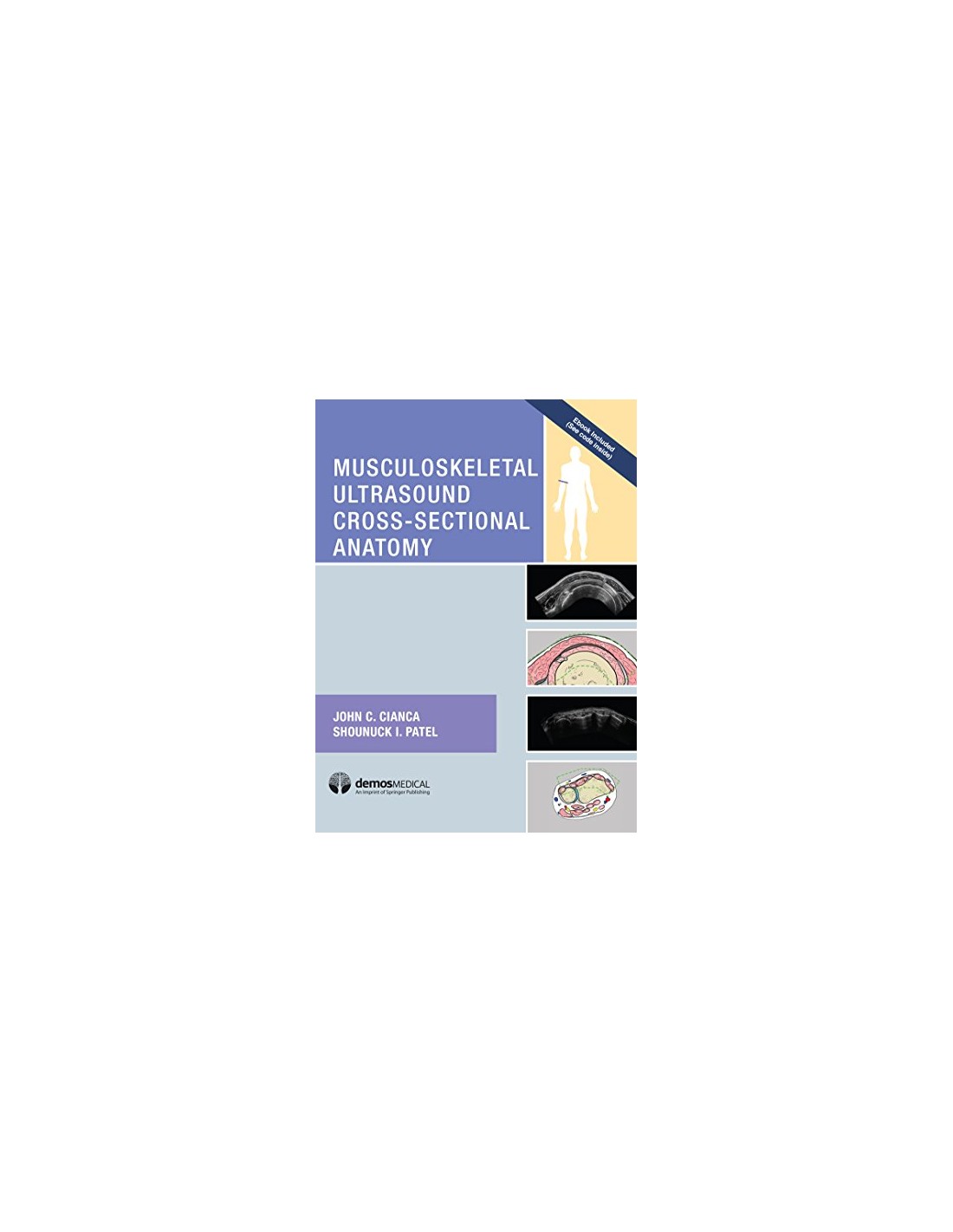
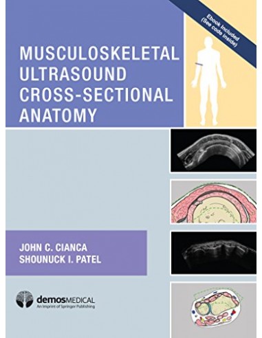
Musculoskeletal Ultrasound Cross-Sectional Anatomy 1/e 2017 (october 2017)
Autori: Cianca, John C., MD; Patel, Shounuck I.
This spectacular cross-sectional atlas provides a roadmap of normal sonographic anatomy of the musculoskeletal system with optimized images that emphasize spatial relationships and three-dimensional orientation.
The book is designed to help novices acquire pattern recognition skills to resolve images into their anatomic components by pairing ultrasound scans with cross-sectional drawings. It will enhance familiarity with musculoskeletal anatomy as it appears on ultrasound imaging for practitioners at any level. Using a sectioned approach, the authors present a visual baseline for evaluating tendon, muscle, ligament, and nerve problems in the upper extremity, lower extremity, and cervical regions. Multiple high resolution views of each structure are accompanied by original illustrations that document the structures in the sonograph and serve as a reference to decipher the image and foster understanding of anatomic relationships and ultrasound appearances.The atlas is an indispensable tool for clinicians learning diagnostic ultrasound, as they can use the anatomical images for comparisons with their own scans. For the seasoned practitioner, the organized format with high-resolution examples makes this an essential reference for confirming exam findings.
KEY FEATURES
- Orients users to anatomical relationships best seen in cross section and necessary to effective utilization of diagnostic ultrasound
- Over 150 ultrasound images cover musculoskeletal anatomy from the shoulder to the foot
- Each ultrasound image has a correlative drawing in cross-sectional or regional format with the scanned area depicted within a highlighted frame to enhance understanding of the scanned anatomy
- Each image is accompanied by a body icon illustrating the level of the scan for each region
- Brief text points and legends emphasize key features and landmarks and offer technical tips for obtaining and interpreting scans
- Autori
- Cianca J C,Patel D O
- Anno
- 2017
- Edizione
- 01
- Volume
- 1
- Pagine
- 380
- Formato
- 22x28
- Aspetto
- Cartonato
9781620700624
Nuovo prodotto
Nessuna recensione
Seleziona per lo zoom